


Presentation
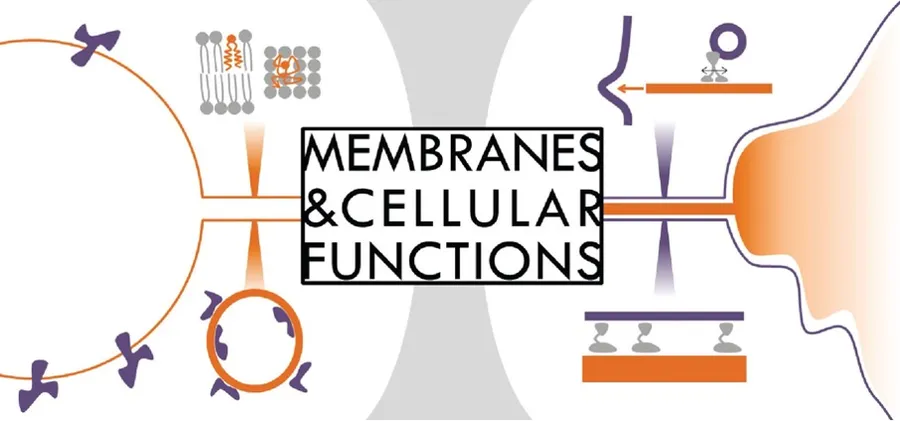
Lipid membranes exhibit non-trivial properties especially at length scales larger than protein molecular sizes. A purely molecular description of membranes is insufficient to arrive at a quantitative understanding of their functions and require meso-scale concepts coming from soft matter and statistical physics.
In addition, cell membranes involve a wide number of membrane-interacting proteins that can alter the overall physical descriptions of the membrane itself. Our goal is to contribute to a more comprehensive understanding of biological membranes and their role in living systems.

Model bio-membranes and cell membranes
To understand the role of lipid membranes and associated proteins involved in essential cellular functions such as transmembrane transport, protein diffusion in membranes, intracellular trafficking, endo/exocytosis, adhesion, cell infection or cell-cell communication, our group develops multidisciplinary approaches that are largely based on synthetic biology, biomimetic systems and quantitative physical measurements. The team has successfully developed several physical approaches for the micromanipulation of giant unilamellar vesicles (GUVs) and cells, combining micropipette aspiration and optical tweezers with confocal microscopy. These technological approaches are particularly powerful for the study of membrane mechanics, both with GUVs and cells, and for studying the roles of membrane curvature and tension. In addition, we use single molecule techniques to study the behaviors of single proteins in membranes. Our research is motivated by close collaborations with biologists and theoreticians, both within and outside Institut Curie.
Members






Publications
Nature Communications - 31/01/2025
See authors
Science Advances - 14/10/2022
See authors
Nature Communications - 01/12/2020
See authors
eLife - 20/09/2018
See authors
Life of the team
Our projects
Membrane trafficking: sorting, bending, cutting

membrane trafficking. Illustration Joanna Podkalicka
Our lab also studies membrane trafficking within the cell and the mechanisms controlling membranes flux in and out of the cell by processes like endo- and exocytosis. Our interest focuses on both proteins and lipids involved, and we are particularly interested in curvature dependent/mediated phenomena, which play a crucial role during all types of vesicular transport. We address questions how different proteins and lipids contribute to the formation of highly curved membrane compartments and how curvature can control their activity. In addition, we study the mechanisms involved in the last step of vesicle formation: membrane scission.

Project investigators
Joanna Podkalicka, Zhi-Qian Wu, Pulkit Aditya, Feng-Ching Tsai
Subproject 1
A new role of caveolin in sphingomyelin transport to the plasma membrane
Lipid and protein sorting are crucial processes that maintain membrane homeostasis among organelles of eukaryotic cells during intracellular transport providing unique properties of each cellular compartment. We are investigating how lipids such as sphingomyelin, which form stiff membranes, are transported from inner compartments and enriched at the plasma membrane. We address the mechanism of protein-mediated lipid sorting which takes advantage of proteins having affinity for both curved membranes and specific lipid species. The goal of the project is to determine the role of caveolin as a potential protein taking part in SM trafficking to the plasma membrane.
Main Collaborators:
Christophe Lamaze (UMR3666)
Daniel Lévy (UMR168)
Franck Perez (UMR144)
Christopher Burd (Yale School of Medicine, US)
Subproject 2
Membrane scission in cells
Project description
Membrane scission is essential in many cellular processes, including cell division and in the generation of transport vesicles. Right before scission occurs, the membrane of a vesicular bud or a thin tubule is highly curved with a saddle-like neck. Also, proteins are assembled at the pre-scission site to provide mechanical forces to overcome the viscoelastic resistance of the membrane. We are interested in how protein assemblies at pre-scission sites drive membrane scission in different cellular processes, in particular the actin cytoskeleton and ESCRT-III complexes.
ESCRT complex proteins are evolutionarily conserved protein that are involved in numerous membrane remodeling processes like cell division, Multi-vesicular body (MVB) formation, nuclear membrane repair. ESCRT-III complex is recruited by the cell during the last part of remodeling process to perform scission of the membrane. ESCRT-III complex has been shown to have be primarily recruited on membrane with negative Gaussian curvature.
The actin cytoskeleton, more specifically the branched actin network mediated by Arp2/3 complex, has been identified as one of the key machineries involved in membrane scission at the plasma membrane and on many organelles (the trans-Golgi network, endosomes and melanosomes). However, despite its presence, how actin operates membrane scission is largely unknown.
Here, we use in vitro reconstitution systems composed of model membranes and purified proteins, combined with biophysical tools to investigate how the ESCRT III complex and the actin cytoskeleton drive membrane scission.
(A) (B)

scission caused by (A) ESCRT-III and (B) the actin cytoskeleton.

and CHMP2A-ΔC(+CHMP3) inside the tube neck. Scale bar: 5 μm.

induced by CHMP4B-ΔC+CHMP2B-ΔC. Scale bar: 50 nm.
Main collaborators
Christophe Le Clainche (Institut de Biologie Intégrative de la Cellule, I2BC)
Ya-Wen Liu (National Taiwan University College of Medicine, Taipei, Taiwan)
Senthil Arumugam (Monash University, Melbourne, Australia)
Cédric Delevoye - UMR 144)
Bassam Hajj (UMR168)
Aurélie Bertin (UMR168)
Winfried Weissenhorn (IBS, Grenoble)
Pierre Sens (UMR168)
How cells interact with their environment
Cells utilize membrane protrusions such as filopodia and tunneling nanotubes (TNTs), for instance for their migration and to allow cell-cell communication, respectively. These cellular protrusions are very thin membrane structures filled with actin filaments. Here, we aim to reveal molecular and physical mechanisms underlying the formation of filopodia and TNTs using biophysical tools to investigate both in vitro and in cellulo systems. To this end, we have developed an in vitro reconstitution system composed of giant unilamellar vesicles (GUVs) and purified proteins that appear key in the formation of filopodia. Furthermore, using optical tweezers to pull membrane nanotubes out of the cell plasma membrane, we are examining in cellulo the conditions for actin polymerization in tubular geometries mimicking those found in these protrusions.

Sub-project 2
Cell-cell adhesion
Project description
Cells adhere to substrates or other cells not only to maintain physical cohesion, but also to receive information about their microenvironment. A number of adhesion proteins orchestrate this mechano-chemical communication. Here, we study they are involved in the dynamics of the first stages of cell adhesion.

Main collaborators
Chiara Zurzolo (Institut Pasteur)
Stéphanie Descroix (UMR168, IPGG)
Pekka Lappalainen (University of Helsinki), Finland)
Gregory A. Voth (University of Chicago, USA)
Christophe Le Clainche (Institut de Biologie Intégrative de la Cellule, I2BC)
Guy Tran Van Nhieu (Laboratory of Biology and Applied Pharmacology, Universite Paris-Saclay)
Septins
Septins are cytoskeletal filaments that interact with the inner plasma membranes. Septins are ubiquitous in eukaryotes, multi-tasking proteins essential for cell division, cell motility, neuro morphogenesis and a variety of membrane remodeling processes. Septins have an affinity for micrometric membrane curvatures at constriction sites of dividing cells, at the base of cilia and dendrites. Linear filaments of septins, self-assembled from palindromic heteromeric complexes, can further form higher-ordered structures. In addition, septins function in concert with other cytoskeletal partners (actin and microtubules) in a finely tuned interplay. We aim at understanding how septins interact with biomimetic model membranes and thus generate membrane reshaping. This is a collaborative project within three teams at PCC (MMM (A. Bertin), LOCCO (B. Hajj) and S. Mangenot (Université de Paris). We have investigated the curvature-sensing properties of septins using in vitro reconstituted systems with model membranes (Beber et al., Nat. Commun., 2019). We have shown that the specific curvature sensitivity of septins drives membrane reshaping. In addition, we are also studying the potential role of septins as a diffusion barrier.


Biophysics of membrane proteins with single-molecule resolution
Researchers: Kémil Belhadji, Johanna Gerstenecker, Alicia Damm, Ranjit Shiva Gulvady, Raju Regmi
Sub-project 1: Interplay of membrane curvature and transmembrane proteins
Transmembrane proteins are tightly embedded in fluid lipid bilayers where they diffuse laterally. Many membrane proteins are involved in the transport of ions or molecules through the membranes using different sources of energy (ATP hydrolysis, voltage, light, etc) that allow for conformational changes. How mechanical stresses on the membrane can affect these conformational changes and thus protein activity is still an open question. We investigate the feedback between the physical properties of membranes, the functional conformational dynamics of membrane proteins and their diffusion. We perform single molecule experiments on membrane proteins reconstituted in liposomes to study the effect of membrane curvature on the conformations of the protein.
Sub-project 2: DNA-FRET sensors to investigate protein clustering mechanisms
Membrane proteins play a crucial role in various biological phenomena such as endocytosis and signal transduction. These phenomena are heavily dependent on the clustering of membrane proteins. While the propensity of proteins to cluster on the cell membrane has been well established, the physical mechanisms that govern this behavior are far from clear. We combine elements from biophysics, cellular biology and biochemistry, to design a single-molecule FRET-based DNA nanosensor to measure the forces responsible for the clustering of membrane proteins.
Main collaborators
Daniel Levy (UMR 168)
Bassam Hajj (UMR 168)
Emmanuel Margeat (CBS, Montpellier)
Ludger Johannes (UMR3666/U1143)
John Ispen (Univ. Southern Denmark, Odense, Denmark)
Weria Pezeshkian (University of Groningen, Netherlands)
Our protocols
Pulling Membrane Nanotubes from GUVs
Coline Prévost*1,2,3, Feng-Ching Tsai*1,4, Patricia Bassereau1,4, Mijo Simunovic1,5
1Laboratoire Physico Chimie Curie, Institut Curie, PSL Research University, CNRS UMR168, 2Department of Genetics and Complex Diseases, T. H. Chan School of Public Health, Harvard Medical School, 3Department of Cell Biology, Harvard Medical School, 4Sorbonne Universités, UPMC University Paris 06, 5Center for Studies in Physics and Biology, The Rockefeller University
* These authors contributed equally
Please, click on the following link to see the video of this work.
Many proteins in the cell sense and induce membrane curvature. We describe a method to pull membrane nanotubes from lipid vesicles to study the interaction of proteins or any curvature-active molecule with curved membranes in vitro.
Proteo-GUV preparation
Matthias Garten1, Sophie Aimon2, Patricia Bassereau1, Gilman E. S. Toombes3
1Institut Curie, Centre de Recherche, CNRS, UMR 168, PhysicoChimie Curie, Université Pierre et Marie Curie, 2Kavli Institute for Brain and Mind, University of California, San Diego, 3Molecular Physiology and Biophysics Section, National Institute for Neurological Disorders and Stroke, National Institute of Health
Please, click on the following link to see the video of this work.
The reconstitution of the transmembrane protein, KvAP, into giant unilamellar vesicles (GUVs) is demonstrated for two dehydration-rehydration methods — electroformation, and gel-assisted swelling. In both methods, small unilamellar vesicles containing the protein are fused together to form GUVs that can then be studied by fluorescence microscopy and patch-clamp electrophysiology.
Join us
Join Us
Open positions
We are continuously looking for Post-docs, PhD students and Master interns. Please get in touch with us via email.
Current openings
Postdoc: In vitro Reconstitution of Cellular Protrusions
PhD: How do cells use surface topography to migrate without adhering?
Alumni
coming soon


Scientific events
Speakers : Anand Srivastava
Speakers : Danielle L. Schmitt
Speakers : Thorsten Hugel


